ADM - Advanced digital microscopy
GINYS-IRB-002
Julien Colombelli
CORE FACILITY MANAGER
Advanced Digital Microscopy platform is a central unit of IRB Barcelona.
It opened in January 2009 and offers 24/7 access and support to 12 state-of-the-art and conventional imaging instruments for life science research, from high content imaging to microscopy wide field, confocal/multiphoton and super resolution, to emerging techniques. for cell manipulation and light sheet-based 3D imaging.
Access to the ADM service page on the IRB Barcelona website.
Services
Services for IRB Barcelona and external researchers (public or private)
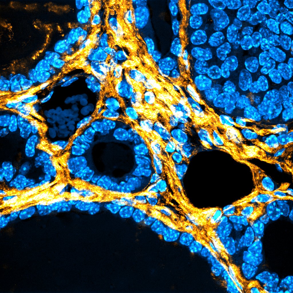
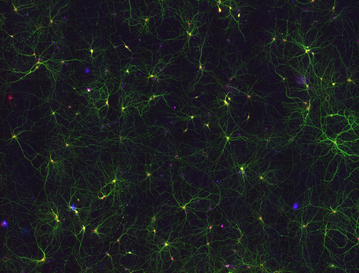
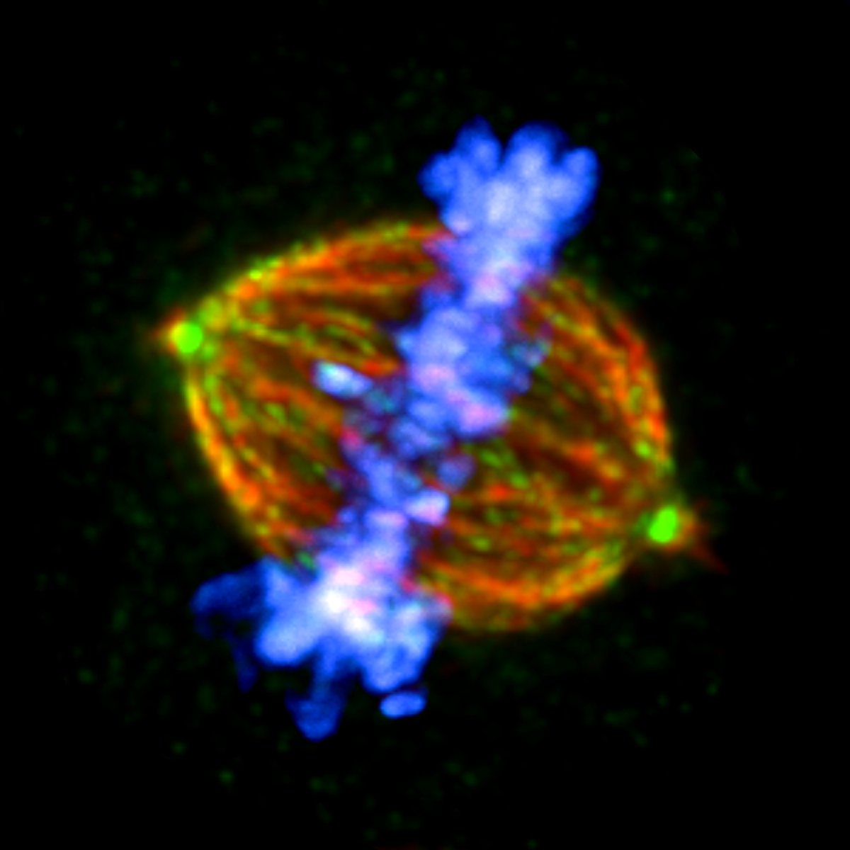
- Spectral laser scanning confocal microscopy. 3D, 4D, and 5D imaging with optical sectioning, custom spectral detection and resolution, high content or multiple position mode, and incubated environment control for live cells.
Four LSM confocal units: inverted confocal systems (Leica SP5, LSM 780, LSM880) and one vertical confocal system (Leica SPE). - Multiphoton microscopy. Deep tissue fluorescence imaging using multiphoton excitations and derived applications (SHG, etc…). Designed in the SP5 with a MAITAI ultrashort powder IR laser (710-990nm).
- Spinning disk confocal microscopy. Fast 5D imaging with the Andor Revolution system, equipped with EMCCD camera technology, FRAP photoactivation and microinjection.
- Multi-Mode Super Resolution System by SIM/ISM/SMLM: The Zeiss Elyra PS1 system combines confocal microscopy (LSM880) with three super resolution modalities: Image Scanning Microscopy (also known as Airyscan) , structured illumination microscopy (SIM), and single molecule localization microscopy (SMLM: STORM, PALM, and variants).
- High-content imaging/detectiont with automated wide-field fluorescence microscopy, confocal spinning disk, or TIRF:
The Olympus ScanR enables a fully automated inverted microscope for wide imaging, multipouc and multiposition plates, automated live imaging with temperature incubation and CO2 control, super-stable illumination for high-quality, reproducible image analysis. content and total internal reflection (TIRF) microscopy for high-contrast images. on the surfaces.
The Nikon LIPSI system enables fast, high-throughput screening imaging with the confocal spinning disk modality (Yokogawa CSU-W1) and multiple multi-well plate readings, thanks to the integrated multi-sample incubator that load samples automatically. The HTS can be performed on up to 20 plates. The system is also well suited for very long-term imaging (duration 1-2 weeks) as it allows robot-driven loading of multiple experiments in parallel. - Laser manipulation of living cells and organisms. FRAP and laser nanosurgery combined in a wide-field fluorescence microscope to perform FRAP and fluorescence patterning, cell ablation, DNA damage, subcellular ablation, and correlative microscopy.
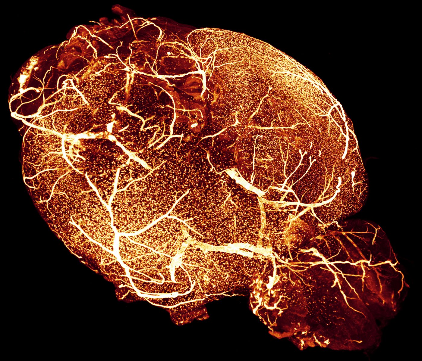
The Zeiss Axiovert 200M laser cutter is a custom platform equipped with a pulsed UV laser (355nm, 470ps) and rotating disc detection. - Light sheet microscopy for optically cleaned organs: a double-sided macroscope-based detection system and double-sided illumination for fluorescence-free 3D imaging and labeling of tissues/organs/organisms that they range from 0.5 mm to 2.5 cm.
- Optical Cleaning: We offer expertise with the latest optical tissue cleaning techniques such as BABB (Murray’s clear), CUBIC, DISCO family, ECI, DeepClear and more.
Lightsheet Microscope based on LEGO bricks, LEMOLISH (http://legolish.org/lemolish) an open source instrument that allows lightsheet imaging of cleaned organs up to 5 cm in size. - Light sheet microscopy for living organisms: a high-resolution double-sided illumination SPIM system, including multiple views and sample rotation, for full-volume live imaging, for samples ranging from 50 µm to 1 mm.
- Light Sheet-Based Oblique Plane Microscope: A light sheet-based system customized on an inverted microscope to enable imaging of multi-well plates and other conventional sample carriers similar to an plate.
- High Throughput OPM. A highly automated OPM system for high-content imaging of organoids in multiwell plates, part of the AECC-funded MACH3CANCER (https://mach3cancer.org) project.
Equipment
The ADM service is committed to developing research instrumentation to adapt, enhance or newly implement innovative biology-driven imaging applications, for example in the field of mesoscopic imaging ( light sheet-based fluorescence microscopy).
The facility offers 24/7 full-time access at:
- Conventional transmission and epifluorescence microscopy and motorized fluorescence macroscopy<
- Spectral Confocal Microscopy, including FRAP, FRET, Time Related Photon Counting (TCSPC) Fluorescence Lifetime (FLIM), Photoactivation and Multiphoton Microscopy
- Spinning disk confocal microscopy
- TIRF microscopy
- Automated high-content imaging microscopy equipped with epifluorescence
- High content protection’ equipped with robotic loader and rotating disk
- Laser nanosurgery of living cells and organisms combined with FRAP
- Optical Clean Light Sheet-Based Whole Organ Imaging
- SPIM based on Lightsheet for living organisms
- Image processing workstations, including HIVE (Acquifer) storage with 240 TB and compute units (as of 2021)
- Light sheet-based oblique plane microscopy for large samples to the inverted microscope
Projects
Title:
NEUBIAS - A New Network for BioImage Analysts
Reference:
http://eubias.org/NEUBIAS
Funding Organism:
COST-UE
Status:
Executing
Publications
Innovating in a bioimaging core through instrument development
Journal Of Microscopy (2024)
Journal Of Microscopy (2024)
Urease-powered nanobots for radionuclide bladder cancer therapy
Nature Nanotechnology 19 554 - 564 (2024)
Nature Nanotechnology 19 554 - 564 (2024)
Expansion microscopy of the chick embryo neural tube to overcome molecular crowding at the centrosomes-cilia
Star Protocols 4 (1 ) 101997 (2023)
Star Protocols 4 (1 ) 101997 (2023)
Neurogenesis redirects β-catenin from adherens junctions to the nucleus to promote axonal growth
Development 150 (16 ) dev201651 (2023)
Development 150 (16 ) dev201651 (2023)
The P4-ATPase Drs2 interacts with and stabilizes the multisubunit tethering complex TRAPPIII in yeast
Embo Reports 24 (5 ) e56134 (2023)
Embo Reports 24 (5 ) e56134 (2023)
Transient and DNA-free in vivo CRISPR/Cas9 genome editing for flexible modeling of endometrial carcinogenesis
Cancer Communications 43 (5 ) 620 - 624 (2023)
Cancer Communications 43 (5 ) 620 - 624 (2023)
Metastatic recurrence in colorectal cancer arises from residual EMP1+ cells
Nature 611 (7936 ) 603 - 613 (2022)
Nature 611 (7936 ) 603 - 613 (2022)
Targeted immunotherapy against distinct cancer-associated fibroblasts overcomes treatment resistance in refractory HER2+ breast tumors
Nature Communications 13 (1 ) 5310 (2022)
Nature Communications 13 (1 ) 5310 (2022)
The LEGO® brick road to open science and biotechnology
Trends In Biotechnology 40 (9 ) 1073 - 1087 (2022)
Trends In Biotechnology 40 (9 ) 1073 - 1087 (2022)
Control of hormone-driven organ disassembly by ECM remodeling and Yorkie-dependent apoptosis
Current Biology 31 (23 ) 5261 (2021)
Current Biology 31 (23 ) 5261 (2021)