SLN - Super resolution Light Microscopy and nanoscopy facility
GINYS-ICFO-001
Dr. Pablo Loza-Alvarez
SLN Head
The Super-resolution Light Microscopy & Nanoscopy (SLN) Research Facility at ICFO is equipped with front-end microscopy techniques that are able to operate a step beyond the commercial state of the art.
The SLN research team performs continuous R&D in most of the advanced light microscopy techniques and provide access and training to all types of users in the forefront of microscopy for the most demanding biomedical applications. The SLN is open to external collaborations with industry, hospitals, research centers and Universities.
The SLN Facility at ICFO provides:
Access to external researchers and collaborators to the variety of state-of-the-art microscopy and super resolution techniques.
Training, through short-term and mid-term, hands-on courses tailored to the needs of specific users with a variety of relevant backgrounds.
Image analysis and quantification tools customized for the different techniques.
Services
The SLN Facility at ICFO provides:
-
- Stimulated emission depletion microscopy: Our STED systems allows 3D super-resolution capabilities in up to 3 optical bands (up to 3 depletion lasers). The single molecule detector (SMD HyD) provides a high detection efficiency. Resonant scanners provide fast scanning capabilities up to videorate at 512×512 pixels. The system includes a white light laser source as well as a 405 nm semiconductor laser. Techniques: STED 3D and 3 colors, Confocal, FLIM, Full-hyper spectral (6D) laser scanning confocal, tau-STED, RESCUE, DYMIN, MINFIELD, easy3D
-
- Multimodal microscopy: Our multimodal workstation is built on top of a commercial Nikon Confocal C1-Si. This system contains an additional scanning head and a double filter turret from where a MIRA 900F Ti:Sapphire fs laser is coupled. This allows the simultaneous implementation of multiphoton imaging techniques such as TPEF, SHG/PSHG, THG, as well as the possibility for performing laser surgery or optical tweezers.Techniques: Confocal, hyperspectral confocal, TPEF, 3PEF, SHG, PSHG, THG, Femtosecod nanosurgery,optical tweezers
-
- Raman micro-spectrometer: Our Raman microscope includes two excitation lasers: 785nm and 514nm. The system is fully automatized and allows switching the excitation light and adjust the laser power by using the software Wire. The system includes the capability of auto-alignment and line streaming.Techniques: Confocal Raman spectroscopy, Surface Enhanced Raman Spectroscopy
-
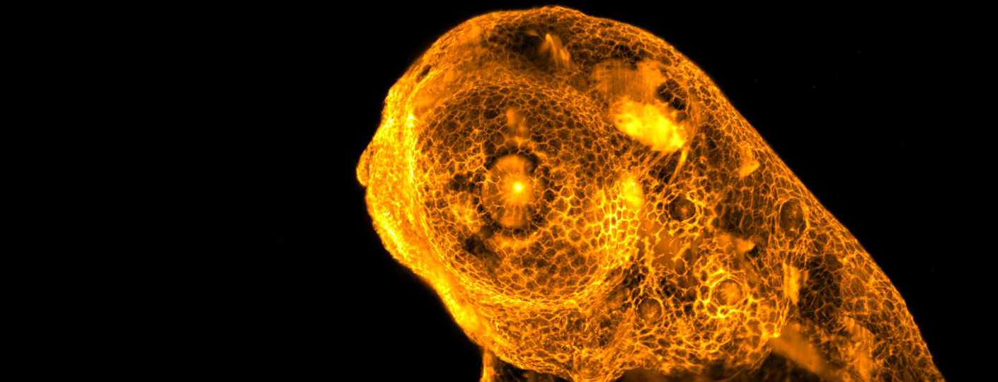
- Light sheet microscopy: Our LSFM systems are custom-made microscopes designed to explore large microscopic 3D samples like organoids or small embryos. They are highly versatile and can be used for adressing diverse biological questions thanks to its modular fashion. They can be set to work with Gaussian and Bessel beams in the linear and non-linear regime. Volumetric images up to 50 vols/s can be achieved.Techniques: TPEF, Digital Scanned Light Sheet Microscopy, Confocal Line Scan, fast volumetric imaging, optical clearing, high throughput, ultramicroscopy.
- Stochastic optical reconstruction microscopy: Our STORM system is based in a TIRF configuration with 4 colors laser bench. The focusing position stability is given by the Perfect Focus System and it is fully motorized. Multicolor Single Molecule Localization acquisitions can be obtained by using the high sensitivity of the EMCCD and 3D Localisation is obtained by introducing the cylindrical lens.Techniques: STORM, PALM, SPT, dSTORM.
Equipment
Leica TCS SP8 STED 3X
Microscope objectives:
10x, 63x and 100x
Excitation wavelengths:
white light laser: 470-670 nm
Semiconductor: 405 nm
8 simultaneous excitation laser lines
STED lasers:
592nm, 660nm, 775nm
Detectors:
2 Backwards HyD’s
1 Forward PMT
2 Backwards PMT’s
Scanning mirrors:
Galvanometric
Resonant
Leica TCS SP5 STED CW
Microscope objectives:
10x, 20x, 40x, 63x and 100x
Excitation wavelengths:
Argon laser: 458, 476, 488, 496, 514 nm
HeNe laser: 543, 633 nm
Ti:Sapphire laser: 750-850nm, pulses duration: 120fs, repetition rate 76MHz
STED lasers:
592nm, CW
Detectors:
2 Backwards HyD’s
1 Forward PMT
3 Backwards PMT’s
2 Backwards APD’s
Scanning mirrors:
Galvanometric
Resonant
Abberior Infinity
Microscope objectives
60x, WD: 0.15 mm, NA=1.42
10x, NA 0.4 air
40x, WD: 0,18 mm, NA=0.95, variable cover glass correction ring
60x silicone oil, WD: 0.3 mm, NA=1.3, variable cover glass correction ring
40X, WD: 0.15 mm, NA=1.3 oil
Universal Plan Extended Apochromat oil immersion objective with 40x magnification
60x, WD: 0.28 mm, NA = 1.2 water
Excitation wavelengths:
Laser Module 405 nm (cw)
Laser Module 485 nm (pulsed, picosecond)
Laser Module 518 nm, (Pulsed, picosecond)
Laser Module 561 nm (pulsed, picosecond)
Laser Module 640 nm (pulsed)
STED lasers:
775nm (high-power) and at 595nm
Detectors:
Spectral detection modules detector
2 single-photon-counting APD modules
2 single-photon-counting optimized for FLIM detection
APD Array detector
Scanning mirrors:
QUAD Beam Scanner: 4 galvo scanners and 4 galvo servo controllers
Nikon Confocal C1-Si
Microscope objectives:
for confocal/multiphoton: 2x, 10x, 20x, 40x, 60x and 100x
specialized for multiphoton (water inmmersion): Olympus 25x , 1.05 NA and Nikon 25x, 1.10 NA
Excitation wavelengths:
Argon laser: 457, 477, 488, 514nm
Diode lasers 405, 561, 635nm
Ti:sapphire 750-850nm
Femtosecond pulsed fiber laser 1550nm
OPO: 1650nm-1750nm
Detectors:
1 Forward multiphoton PMT, Large area IR Photodiode, Vis. photodiode
2 Backwards multiphoton PMT’s
2 Backwards confocal PMT’s and 32 spectral detectors
Scanning mirrors:
Galvanometric
Renishaw InVia Raman micro-spectrometer
Microscope objectives:
5x, 20x long working distance, 20x, 50x and 100x
Excitation wavelengths:
785nm 300mW
532nm 100mW
Light Sheet Fluorescence Microscopes (4 different systems)
Microscope objectives:
for excitation: 10X, 0.3 NA and 10X, 0.45 NA
for collection (water inmmersion): 20X, 0.5 NA and 25X, 1.05 NA
Excitation wavelengths:
Diode lasers 405nm, 488nm, 638nm
DPSS laser 532nm, 651nm
Ti:Sapphire 750-850nm
Femtosecond fiber laser 1030nm
Detectors:
Hamamatsu Orca R2
Hamamatsu Orca Flash 4
Nikon N-STORM microscope
Microscope objectives:
10x, 40x, 60x and 100x
Excitation wavelengths:
405, 488, 561, 647 nm
Detectors
Camera: EMCCD IXON 897
Staff
Dr. Jordi Andilla | jordi.andilla@icfo.eu
| 93 553 4038 - 93 553 4068 - 93 554 2259 | ORCID
| PRC Page
Dr. Mónica Marro Sánchez | monica.marro@icfo.eu
| 93 553 4038 - 93 553 4068 - 93 554 2259 | ORCID
| PRC Page
Projects
Title:
A multiplexed biomimetic imaging platform for assessing single cell plasticity (Plastomics) and scoring of tumour malignancy
Funding Organism:
EU EIC
Status:
Executing
Title:
Advanced Multimodal Photonics Laser Imaging Tool for Urothelial Diagnosis and Endoscopy
Reference:
https://www.amplitude-imaging.com/
Funding Organism:
H2020 EU
Status:
Executing
Title:
Adaptive Retinal Implant Technology for Vision Restoration (iVISION)
Funding Organism:
Fundacio La Caixa (CaixaResearch)
Status:
Executing
Publications
Few-cycle all-fiber supercontinuum laser for ultrabroadband multimodal nonlinear microscopy
Optics Express Vol. 30, Issue 16, pp. 29044-29062 (2022). https://doi.org/10.1364/OE.454726
Optics Express Vol. 30, Issue 16, pp. 29044-29062 (2022). https://doi.org/10.1364/OE.454726
Focus variation due to near infrared laser in a confocal microscope
Microscopy Research & Technique, Volume85, Issue10 October 2022 Pages 3431-3438
Microscopy Research & Technique, Volume85, Issue10 October 2022 Pages 3431-3438
Multi-modal and multi-scale clinical retinal imaging system with pupil and retinal tracking
Sci Rep 12, 9577 (2022)
Sci Rep 12, 9577 (2022)
Hippocampal Neuronal Cultures to Detect and Study New Pathogenic Antibodies Involved in Autoimmune Encephalitis
JoVE, 184, e63829 (2022)
JoVE, 184, e63829 (2022)
Deficiency of the ywhaz gene, involved in neurodevelopmental disorders, alters brain activity and behaviour in zebrafish
Mol. Psychiatry, 1-10 (2022)
Mol. Psychiatry, 1-10 (2022)
Multiple asters organize the yolk microtubule network during dclk2-GFP zebrafish epiboly
Sci Rep 12, 4072 (2022). https://doi.org/10.1038/s41598-022-07747-7
Sci Rep 12, 4072 (2022). https://doi.org/10.1038/s41598-022-07747-7
Human CASPR2 antibodies reversibly alter memory and the CASPR2 protein complex
Annals of Neurology
Annals of Neurology
Novel Non-Invasive Quantification and Imaging of Eumelanin and DHICA Subunit in Skin Lesions by Raman Spectroscopy and MCR Algorithm: Improving Dysplastic Nevi Diagnosis
Cancers 14, 4, 1056 (2022)
Cancers 14, 4, 1056 (2022)
Constitutive Activation of p62/Sequestosome-1-Mediated Proteaphagy Regulates Proteolysis and Impairs Cell Death in Bortezomib-Resistant Mantle Cell Lymphoma
Cancers 14, 4, 923 (2022)
Cancers 14, 4, 923 (2022)
Modular multimodal platform for classical and high throughput light sheet microscopy
Sci Rep 12, 1969 (2022)
Sci Rep 12, 1969 (2022)