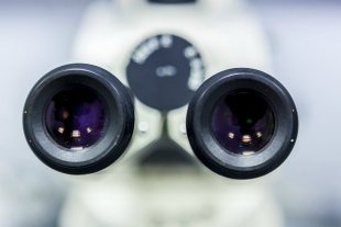
Microscopy
GINYS-IGTP-005
Instal·lació de Microscopis
Institut de Recerca Germans Trias i Pujol (IGTP)
Planta Baixa
Carretera de can Ruti, camí de les escoles, s/n
08916 Badalona
Institut de Recerca Germans Trias i Pujol (IGTP)
Planta Baixa
Carretera de can Ruti, camí de les escoles, s/n
08916 Badalona
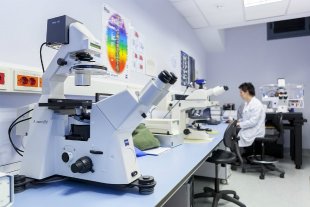
The Microscopy facility was created to provide scientific and technological support for microscopy and related services to research groups and clinical services. The facility provides consultations both IGTP and external groups who require it. There is a dedicated preparation area for samples and a observation area for producing and analysing images.
Training sessions are organized periodically for students and technical staff. There is also a consultation service.
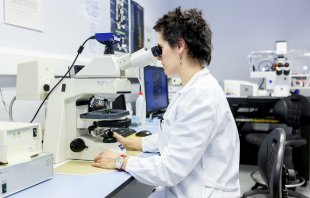
Depending on previous training and skills of users they can use the facility on a self-service basis with the help of a specialist technician.
Services
- Production of cryoblocks, cryosections and cell preparations
- Single or multiple staining and immunocytochemistry production. It is also possible to perform morphometric analyzes of images.
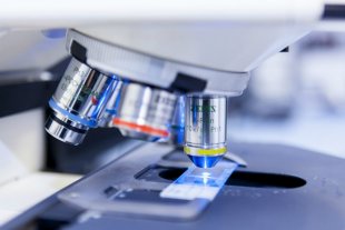
Axioskop 2 microscope (Zeiss) with color digital camera for high resolution images.
- Multiple fluorescent labeling of tissues, cells and sections of biological samples in multiposition, panoramic, Z-axis multipositin and time-lapse
Zeiss, microscope model AxiObserver Z1. The motorized instrument allows to see panoramic views, in time lapse and in multiple positions of objects mounted in slides or in plates or flasks of Petri. The Apotome module for optical sections prevents the scattering of light even in thicker samples giving higher quality images.
- Fluorescence staining images of living cell cultures with CO2 and temperature controlled incubation chamber
The AxiObserver Z1 (Zeiss) has an external culture chamber with controlled CO2 temperature. This allows an incubation of up to 7 days without changing the conditions of the culture. In this case images can be obtained using the Apotome module for better quality.
- Multipocal confocal images, multiposition Z-axis panoramas or time lapse
Zeiss AxiObserver Z1 microscopy coupled to a confocal LSM 710. There are seven laser beams available to allow multiple marking on images.
- Three-dimensional analysis: Z-axis quantification, three-dimensional reconstructions of Z-series confocals, etc.
Complementary Services
Consultation on design, analysis and interpretation of results.
Equipment
- Cryostatic microtome with UVC disinfection and Ag-protect LEICA CM1950 system for handling potentially dangerous specimens. Semi-automatic with 15 plate positions, working temperature range from 18ºC to -42ºC and rapid freezing of samples up to -52ºC and vacuum washing system to reduce direct handling of samples.
- Cytofuge® 2 centrifugation system (StatSpin) for obtaining cellular preparations on slides.
- Zeiss Axioskop 2 light field and epifluorescence microscope with filters for the most common fluorochromes (filter sets 1, 9, 10 and 15) with 5x, 10x, 20x, 40x and 63x oil lenses coupled to an AxioCam MRC5 color camera (D). ZEN blue 2011 software to obtain and analyze images.
- Zeiss AxioObserver Z1 inverted fluorescence microscope with 1, 20 and 38HE filter sets, 10x, 20x, 40x, 40x oil and 63x oil objectives. Attached to the AxioCam MRc5 (D) monochrome camera and the 2012 ZEN blue software to obtain and analyze images. For average living cell length studies, there is an external incubation chamber with a modular heating unit for 6 w and 24 w plates with CO 2 temperature and ambient regulation. To capture images in optical sections there is an Apotome module to prevent light scattering, even in thicker samples.
- Zeiss LSM 710 confocal module coupled to the Zeiss AxioObserver Z1 microscope equipped with 7b laser beams (405, 458-488-514, 543, 594, 633); 10x, 20x, 40x, 40x and 63x oil lenses and ZEN black 2012 software for image acquisition and analysis.