Microscopy
GINYS-IGTP-005
IGTP Microscopy Platform Institut de Recerca Germans Trias i Pujol (IGTP)
Planta Baixa
Carretera de can Ruti, camí de les escoles, s/n
08916 Badalona
Planta Baixa
Carretera de can Ruti, camí de les escoles, s/n
08916 Badalona
The Microscopy Platform was created to provide scientific and technological support to research groups and clinical services for microscopy-based experiments throughout all steps from experiment planning and execution, imaging, to image post-processing. The facility provides consultations both IGTP and external groups who require it.
Initial training and refresh sessions are organized for new and returning users on request. Trained and authorised users can then use the facility on a self-service basis or with the help of the facility staff, depending on their needs.
For all queries please contact Dr Jakub Chojnacki.
Initial training and refresh sessions are organized for new and returning users on request. Trained and authorised users can then use the facility on a self-service basis or with the help of the facility staff, depending on their needs.
For all queries please contact Dr Jakub Chojnacki.
Services
IGTP Microscopy platform provides the following services:
- Automated widefield and confocal microscopy
- Live-cell widefield or confocal imaging
- STED Super-resolution microscopy
- Spectral imaging
- Fluorescence Correlation Spectroscopy (FCS and STED-FCS)
- Image processing and analysis
Complementary services
IGTP Microscopy platform also offers full support and advice for microscopy-based experiment design, execution and data analysis as well training in efficient and responsible use of the microscopy equipment.
Equipment
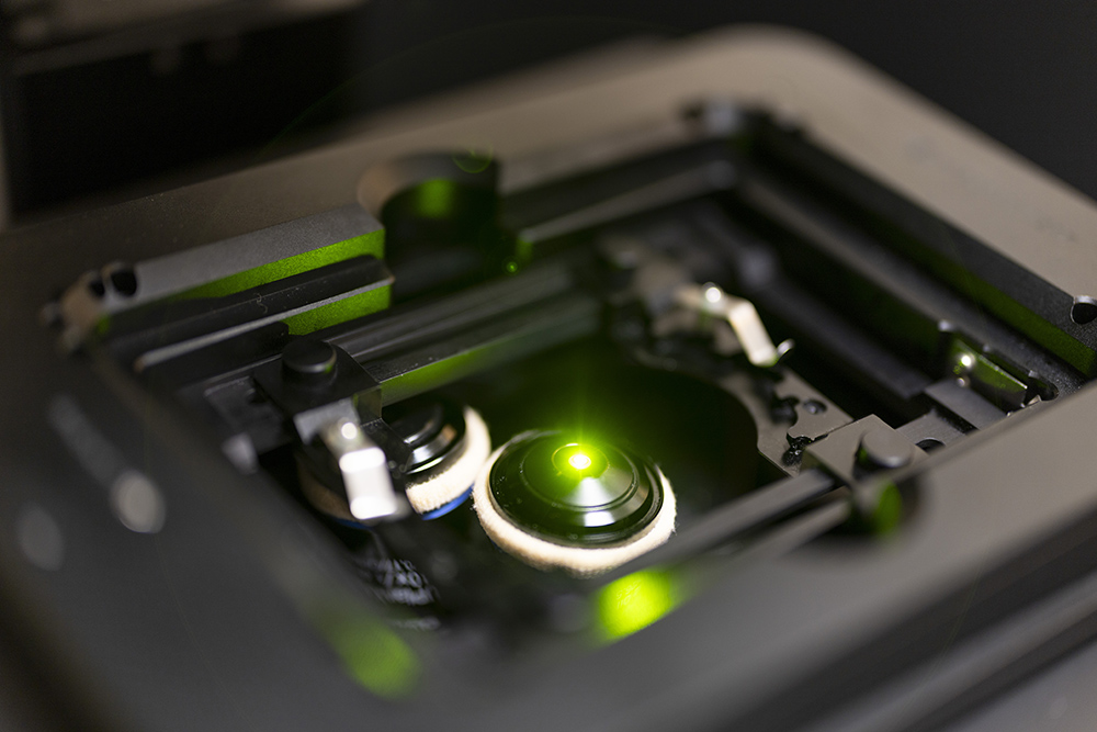
- Abberior INFINITY From May 2023, high-level biosafety (NCB3) facility of the Comparative Medicine and Bioimage Centre of Catalonia (CMCiB) houses Abberior Infinity; a state-of the-art super-resolution confocal microscope. This microscope is adapted for live cell imaging, and thanks to its super-resolution capabilities, is specifically well suited for studying molecular details of pathogen (viruses, bacteria, parasites) interactions with cells, organoids or tissues. Finally, due to open and easily upgradable nature of this platform, it will be able to grow depending on users’ needs and offer in the future additional modalities such as 2-photon excitation or live animal intravital imaging. The equipment of this class, unique in the region, is a powerful tool for advanced microscopy studies on fully infectious Level 3 pathogens (HIV, SARS-CoV-2, Monkey Pox, TB etc.) It is available for use by all internal and external users via IGTP Microscopy Platform.
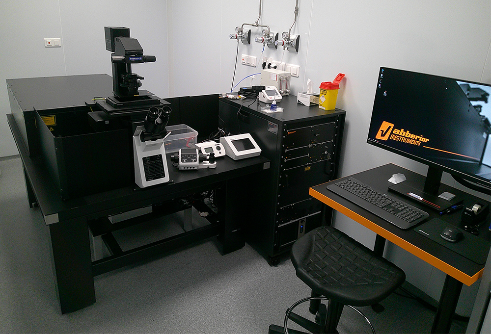
- Abberior STEDYCON This microscope is a compact confocal & super-resolution microscope capable of 4-colour imaging in confocal and STED super resolution modes (resolution down to 40 nm). STEDYCON is coupled to fully motorised Olympus IX83 body equipped with QUAD-band epifluorescence illumination as well as 10x, 20x dry objectives plus 40x and 63x oil objectives. STEDYCON software for obtaining and analysing images also comes with SVI Huygens Pro software for fast image stitching and deconvolution.
- Leica DMI6000B Fully motorised Leica DMI6000 B inverted widefield microscope for brightfield and fluorescence imaging with fluorescence filter sets for UV, Blue and Orange light excited fluorophores as well as 4x, 10x, 20x, 40x dry objectives plus 63x oil objective. The microscope is coupled to Leica DFC420 5MP colour and DFC360 1.4 MP monochrome camera, and utilises Leica LAS X software for obtaining and analysing images.
- Zeiss PALM A microdissection system for non-contact sampling and capture of RNA, DNA and proteins as well as live single cell cloning and culture. This fully motorised system built on Zeiss Axio Observer Z1 inverted microscope is equipped 10x, 20x, 40x dry objectives for brightfield-only imaging and microdissection. Laser Microdissection and Pressure Catapulting (LPMC) is realised via 355 nm UV laser. The microscope is coupled to Hitachi HV-D30 and Zeiss AxioCam MRc5 5 MP colour cameras.
- Zeiss Axio Observer Z1 Inverted widefield microscope for brightfield and epifluorescence imaging with fluorescence filter sets for UV, Blue and Orange light excited fluorophores as well as 10x, 20x, 40x dry objectives plus 40x and 63x oil objectives. The microscope is coupled to Zeiss AxioCam MRm 1.4 MP monochrome and utilises ZEN blue 2012 software for obtaining and analysing images. For live cell imaging studies, the microscope is equipped with an incubation chamber (full enclosure) with a modular heating unit for Petri dishes, microscope slides, and 24-well plates and regulation of temperature and CO2 environment.
- Leica CM1950 cryostat A cryostatic microtome with UVC disinfection and Ag-protect system for the manipulation of potentially dangerous specimens. The cryostat is semi-automatic with 15 plate positions, working temperature range from 18 °C to -42 °C, rapid freezing of samples to -52 °C and vacuum washing system to reduce direct manipulation of samples.
- Leica M80 A stereo microscope for microdissections equipped with LED3000 RL ring illuminator, transmitted light base as well as KL1500 LCD spotlight illumination light source. The microscope is coupled to a stand-alone digital imaging system based on FLEXCAM C1 12 MP colour camera.
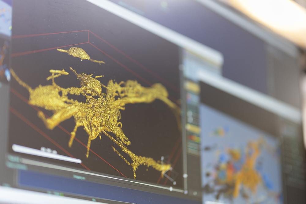
- Image Processing Workstation A dedicated workstation (CPU: 16 core Intel Xeon W5-2465X 4.50GHz, 256GB DDR5 ECC RAM, GPU: NVIDIA RTX A5000 24GB) designed to assist in handling large data sets and complex image processing tasks. It comes with SVI Huygens Professional commercial image processing and visualisation software as well as a number of open-source image processing applications.